 Life Sciences
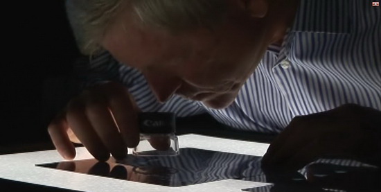
Peering into the Chrysalis: An Interview with Dr. Richard Stringer
The transformation from caterpillar to butterfly inside a chrysalis has been a profound mystery of nature. Using MRI for the first time on a chrysalis, however, Dr. Richard Stringer has begun to reveal the “inside story” of metamorphosis.
 Dr. Stringer, how did you get interested in butterflies?
One day as I watched an elegant Monarch butterfly emerge from its chrysalis l wondered if there could be some way to get inside a chrysalis and watch the caterpillar to adult butterfly transformation process. Later I happened to be looking at MRl slices of a human in an anatomy text book. That work had been done at the Center for In Vivo Microscopy at Duke University Medical Center in August 1998. I immediately called up the people at Duke. They were as curious as I was about what MRl could reveal using their very powerful MRI equipment. They offered to scan the chrysalids over a period of 10 days. I raised some money from a generous person on the Internet.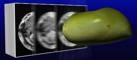
how MRI slices a Monarch chrysalis
into more than 200 images.
Besides the MRI work was there anything else memorable about the trip to have the MRI work done?
During the trip to Durham l stopped at a gas station and someone jumped into my car and drove away with chrysalids, my computer, and research notes. A potential disaster? No! Thanks to another butterfly aficionado and the Internet, I had substitute live chrysalids the next day.
What did you see in the MRI results?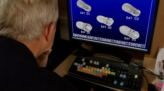
Looking at the resulting MRI slices you can see the differences in development (metamorphosis) as the week progressed. The most obvious changes involved wing development. Incredibly, the butterfly brain could already be seen in the first day chrysalis. The digestive tract changed from being an intestine-like organ in the early chrysalis to reproductive organs in the mature chrysalis.
If you were to label this chrysalis exploration you have done what would the title be?
“There is more than one way to look at a butterfly chrysalis.”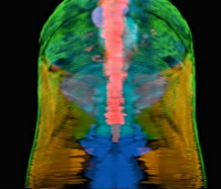
animated Dr. Stringer’s MRI images into
a 3-D composite that allows the viewer
to “fly through” the interior of a chrysalis
as the butterfly organs develop.
How did you link up with Illustra Media?
It was during the early days in formulating information about butterfly chrysalids. Lad Allen, the producer, through one of his contacts, had found a reference to my MRI work. We had several phone conversations before I met with Lad and Jerry Harned at their studio in California where we reviewed all my material and scenarios for using the material in Metamorphosis.
What is your background? First of all, let me say that it is projects like this one that make me thankful that I have had a versatile background. My doctorate in environmental health is from the Johns Hopkins School of Hygiene and Public Health. Following that I worked for EPA as a biologist, and then spent 10 years with an engineering company as a biologist working on everything from Nuclear Power Plants to water quality studies in Egypt. I’ve been the Environmental Director for a Japanese battery manufacturer and presently teach Biology and Environmental Science at Harrisburg Area Community College in Lancaster, Pennsylvania.
Every summer I raise butterflies on my land in Lancaster. The land backs up to a waterfall. It is perfect for raising butterflies and the flowers they need. I use the butterflies for research, for teaching, and profit. But really mostly, I raise the butterflies for the smiles they bring.
What do you like most about butterflies?
They make everyone smile.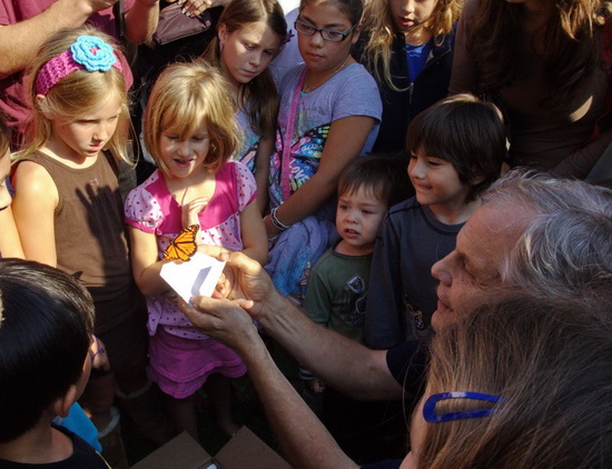
Besides the MRI work, have you used other technologies to watch the butterfly chrysalis as it matures eventually becoming an adult butterfly?
I was really infatuated with the whole metamorphosis process and kept an eye out for new technologies that could be used non-invasively to track changes occurring during the caterpillar to adult butterfly transformation. I molded and poured a foot long bronze chrysalis at a foundry in town. We used mammography X-ray at a local hospital to see what X-ray could reveal about the chrysalis. We used CT (computer aided tomography). We used the CT data to 3-D print the outside and inside of a chrysalis. We also used micro-CT technology and with it produced some of the best images of the study. It selectively revealed the spiracles and tracheal tubes.
Those gold spots encircling the abdomen of the monarch chrysalis: do you know what they are?
That question is one of the first ones asked by people who see a monarch chrysalis. In fact, those gold spots look like real gold and they have served as a model for jewelry. The micro-CT shows that the gold spots are spiracles, points of entry for air entering the chrysalis.
Anything else you’d like to share?
Ask me about how we are using nanotechnology to listen to the butterfly heartbeat…
That would be amazing — to hear a beating heart in a creature weighing a fifth of an ounce! We’ll have to “listen” for that. Thanks very much for sharing your expertise with us.
____________________________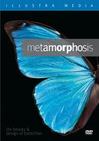 To view Dr. Stringer’s MRI images of the Monarch chryalis developing in exquisite detail, order Illustra’s superb documentary, Metamorphosis, the Beauty and Design of Butterflies. available in both DVD and Blu-Ray formats. While visiting the website, you can download a free copy of the companion e-book, Metamorphosis, the Case for Intelligent Design in a Chrysalis from Discovery Institute Press. And here at ENV, search on Metamorphosis for previous interviews and articles about the production.