 Evolution
Reconstructing Kimberella — The Disputed Anatomy in Detail
Editor’s note: We are delighted to present a series of posts by paleontologist Günter Bechly on the Ediacaran organism Kimberella. If identified as an animal, it would “predate the Cambrian explosion of bilaterian animal phyla as a kind of ‘advance guard.’” The question is of interest for debates about evolution and arguments about intelligent design raised by Stephen Meyer, among others. Find the full series about Kimberella here.
In my opinion the third phase reconstruction of Kimberella, by Fedonkin (2007b) and described here yesterday, is better supported by the fossil evidence than the new reconstruction of Ivantsov (2009). Not only is Fedonkin more likely correct concerning the head and feeding apparatus with a medium sized proboscis armed with two teeth. He also successfully addressed all the arguments that Ivantsov listed against a single shell. You can find a pretty decent life-reconstruction and animation of Kimberella at the PalaeoZoo website.
Foot and Mantle
With their revolutionary new interpretation of Kimberella as a benthic bilaterian animal, Fedonkin & Waggoner (1997) first recognized the presence of a creeping ventral locomotory organ (foot), which was accepted by most subsequent authors (a notable exception was Dzik 2003). Fedonkin et al. (2007b) explicitly identified this structure as “a true foot, possibly comparable in structure with that of monoplacophorans.” Fedonkin et al. (2007b) also described fine transverse wrinkles on the sole of the foot, which they interpreted as transverse ventral musculature (compare Seilacher et al. 2003 who likewise described the “flat foot with a ring of segmental muscles, whose contraction upon death produced equidistant wrinkles”). They suggested backward movement by peristaltic waves and also suggested that the foot was contracted in some specimens in a manner similar to modern limpets.
It was again Fedonkin et al. (2007b) who first explicitly suggested the presence of a mantle and mantle cavity in Kimberella. Ivantsov (2009) agreed, but consistently used the term “mantle” with quotation marks because he doubted a homology with the molluscan mantle or pallium, which is the technical term for their protruding dorsal body wall.
More recent authors all affirmed the presence of a mantle: For example, Gehling et al. (2014: fig. 9) mentioned that “the serrate zone has been interpreted as a corrugated mantle frill overlying a wider central muscular foot.”
According to Wanninger & Wollensen (2019) Kimberella has an “elongated, slender foot surrounded by a mantle that is separated from the former by a circumpedal mantle cavity” (also see Vinther 2015).
In the most recent textbook on invertebrate phylogeny, by Giribet & Edgecombe (2020), the authors say about Kimberella‘s anatomy that “the softer ventral side has been described as a sole and compared to a molluscan foot. The body is certainly zoned laterally, this zonation viewed as representing the foot and mantle separated by a groove in the mollusc model.”
It is safe to say that since the redescription of Kimberella as a benthic bilaterian animal, the functional interpretation of foot and mantle has met with a broad consensus among the specialists and represents one of the less controversial issues of its anatomy. However, what remains highly disputed is the question of the homology of these structures with the foot and mantle in modern mollusks.
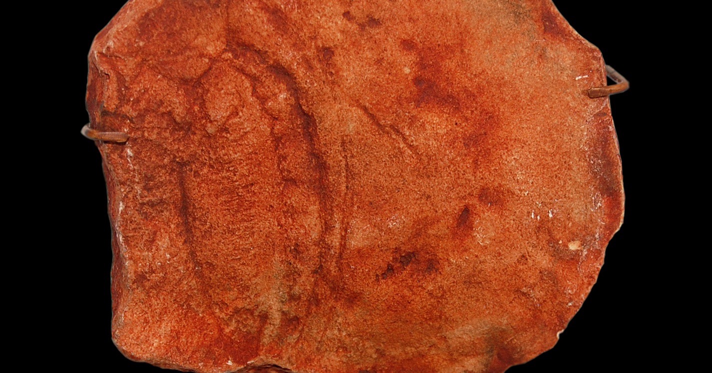
Shell
According to esteemed Russian expert Mikhail Fedonkin, Kimberella had a non-mineralized univalved shell with an elongated oval, shield-like outline (Fedonkin & Waggoner 1997, Fedonkin 2001, 2003, Fedonkin et al. 2007b). Likewise, Fedonkin (1998, 2001) and Waggoner (1998) said that Kimberella “bore a highly compaction-resistant structure that we interpret as a stiff but un-mineralized shell,” and Fedonkin (2003)specified that the “elongated and high dorsal shell [was] made of flexible and rigid organic material.” Fedonkin et al. (2007b) found that “the outer surface of the dorsal side of the smaller specimens is covered with numerous round protuberances, uniformly spaced over the major part of the shell,” which they interpreted as outgrowths, or bases of mineral spines, or as “separate initial nodules of shell formation.” This latter hypothesis might explain why these structures are not visible in the larger adult specimens. Finally, Fedonkin et al. (2007b) also recognized that small specimens often are strongly elongated, and suggested that “these individuals represent a phase when the formation of the shell had not yet begun or was just incipient.” They explained that “the taphonomic varieties of the shell imprints clearly demonstrate that the shell was stiff, but thin and flexible, particularly in juveniles,” which explains why stretched and laterally bent specimens are generally small.
Based on further new material, the other Russian expert Andrey Ivantsov (2009, 2010b) suggested a new reconstruction with “hard sclerites, probably of aragonite, … rather than a complete consolidated shell.” Because of the very different contracted and stretched morphs of Kimberella, Ivantsov (2009: pls I-II, 2010b: pl. 1, 2013) claimed that a rigid shell was not a possibility, and instead suggested a soft-bodied and extremely flexible organism with multiple dorsal mineralized sclerites. He said that “many characters of the imprints contradict the hypothesis of a single shell,” including the many stretched and strongly bent specimens. This is somewhat strange, because we have just seen that Fedonkin et al. (2007b) had already explained this phenomenon ontogenetically. Ivantsov also claimed that the absence of any growth lines is very much unlike molluscan shells, even though Fedonkin et al. (2007b) suggested that “the absence of any growth zonation in the shell structure suggests that the shell of Kimberella was the homologue of the periostracum of later Mollusca.” Again and again we find opposite conclusions about the same evidence. Finally, Ivantsov mentioned that the head possessed tubercles similar to the body and claimed that all “these considerations leave no doubt that Kimberella had no single dense shell.” Ivantsov (2017) suggested that the dorsal body cover was “armored with fine sclerites, apparently mineral, but rapidly dissolving after burial,” but this taphonomic hypothesis does not explain why the tubercles are only visible in small specimens.
Most later authors followed the single shell interpretation of Fedonkin: Seilacher et al. (2003) mentioned that the “dorsal shield, … was soft enough to become deformed during burial.” Scheltema & Schander (2006) said that Kimberella had a “single, stiff, unmineralized dorsal exoskeleton,” which was formed by the mantle cuticle alone, “although there may have been a zone of spines beneath the dorsum.” Gehling et al. (2014) featured a reconstruction of Kimberella as a molluscan grade organism with a single shell and said that “this reconstruction of Kimberella includes an inferred stiff but flexible unmineralized shield that enclosed internal organs.” Vinther (2015) remarked that Kimberella had a dorsal “cuticular shield with tubercular nodes.” Finally, Wanninger & Wollesen (2019) claimed that the presence of a single non-mineralized shell in Kimberella fits well with the hypothetical ground plan of mollusks, since the discovery of the Ordovician fossil Calvapilosa suggested that the two-shelled state of halkieriids and the multi-shelled state of polyplacophoran mollusks represents a derived condition.
Nevertheless, some later authors rather agreed with Ivantsov’s new interpretation. For example, Seilacher & Hagadorn (2010) concurred that “Kimberella lacked a rigid shell, while being covered by small sclerodermites” (Bengtson 2005, Fedonkin et al. 2007b, Ivantsov, 2009).” Even more recently, Parkhaev (2017) concluded that “in contrast to the first reconstructions …, it was shown that Kimberella had no firm and solid shell, otherwise it would be difficult to explain the observed variability in the shape and proportions of the body in known specimens.”
Lay people must be left quite confused by this controversy and might even conclude that paleontology often seems to boil down to an esoteric exercise in “reading tea leaves.” Unfortunately, sometimes this is the case indeed. Nevertheless, one should always make an inference to the best explanation. In my view the original interpretation of Fedonkin is better supported and he also sufficiently explained the evidence that allegedly supports the alternative interpretation of Ivantsov. Therefore, I conclude that Kimberella more likely possessed a non-mineralized integument, a quasi-leathery shell with a characteristic ornamentation, which was flexible in juvenile specimens but rather stiff in adult ones. But this is just my humble opinion and it is quite clear that this issue is controversial and far from settled.
Gills
Fedonkin & Waggoner (1997) speculated that the crenellated structures may have had a respiratory function and even mentioned the possibility that the crenellations may have hosted microbial symbionts. Fedonkin et al. (2007b) said that “there is no evidence of gills in Kimberella, while the large surface of the multifolded crenulated zone could effectively perform the respiration function.” Fedonkin considered these circumpedal respiratorial folds as “possible predecessor of the ctenidia” (meaning, feathery gills) in aquatic mollusks.
Even though there are indeed no traces of respiratory organs preserved in any of the Kimberella fossils, such gills were proposed by Ivantsov (2009, 2012) as possibly attached to the scallops along the frilled margin, similar to the serially repeated gills in the Cambrian stem mollusk Odontogriphus (Butterfield 2006) and in the circumpedal mantle cavity of primitive living mollusks (Serialia) (Stöger et al. 2013). However, Ivantsov also mentioned the alternative possibility that gas exchange occurred across the general body surface. Likewise, Seilacher & Hagadorn (2010) concurred with Fedonkin et al. (2007b) and Trusler et al. (2007) that “the serial impressions between the foot and the cap probably correspond to flap- or gill-like structures … as in modern chitons and Neopilina, and in the Cambrian Odontogriphus from the Burgess Shale.”
However, all this is of course mere speculation in the absence of any conclusive empirical evidence.
Digestive System
Dzik (2003) mentioned that “the most prominent aspect of Kimberella fossils is a voluminous depression in their center” and proposed that “the most likely interpretation is that this was a gut content.” This median groove indeed was generally interpreted as simple digestive tract and pharynx (Fedonkin et al. 2007b, Knoll 2011, Vinther 2015). A notable exception was Seilacher (1999), who thought that “the deep cleft along the midline of the foot is clearly a secondary feature” related to the burial process and fossilization as ventral death mask (Gehling 1999). Gehling et al. (2014) concurred that the median keel or grove, which is most prominent in smaller specimens, most likely corresponds to a “flexible, unmineralized outer integument.” Déjà-vu, anyone?
Feeding Apparatus
Many authors have suggested that the tapered end of Kimberella corresponded to a kind of proboscis as a feeding apparatus. The alignment between the feeding traces and the tapered “open” end of the body with the assumed proboscis clearly suggests that this was the anterior (oral) end with the pharynx.
Based on the trace fossil evidence, Ivantsov & Fedonkin (2001b) and Fedonkin (2001, 2003) first speculated that the “feeding tracks may reflect the work of the proboscis that bears the hook-like organs on its end.” Fedonkin (2003) thought that a comparison of the body size of Kimberella and of the size and pattern of the feeding traces suggests that the hypothetical proboscis could be extended as long as the whole body of Kimberella, and that there were two sharp teeth at its end. Seilacher et al. (2003, 2005) also speculated that Kimberella was a stationary grazer that repeatedly fed with a long proboscis. Dzik (2003) mentioned that “in all well preserved specimens of Kimberella there is a distinctly delimited narrower part of the proposed gut, perhaps representing a muscular oesophagus or even evertible proboscis.”
Gehling (1996) and Gehling et al. (2005: fig. 12) were the first to clearly document actual fossil evidence for such an extensible head or everted proboscis at the anterior end of the Kimberella animal. Years later, Gehling et al. (2014: fig. 7) featured more specimens with an everted proboscis, which is never longer than about a fourth of the remaining body length, thus not as long as Seilacher & Hagadorn (2010) and others had speculated. The fact that in most specimens this proboscis is not visible was inferred by these authors “to be a result of its retraction within the circumference of the body,” but this is completely ad hoc.
This proboscis was described in detail by Fedonkin et al. (2007b), who also described a pair of bag-like structures lateral at the base of the proboscis (and thus the assumed pharynx) at the anterior end of the body, which they interpreted as oesophageal pouches or “pharyngeal glands (comparable with those found in many extant molluscs)” (Fedonkin et al. 2007b: fig. 15; also see: Ivantsov 2009: pl. 1 figs 7-8, Vinther et al. 2012, Vinther 2015: fig. 3E). Fedonkin suggested that the feeding traces were “left by a solitary conjugate pair of teeth located at the end of a long proboscis that stretched far forward beyond the main body” (Ivantsov & Fedonkin 2001b, Fedonkin 2003, Fedonkin & Vickers-Rich 2007, Fedonkin et al. 2007b).
In the paper by Fedonkin et al. (2007b) an alternative interpretation by co-author Ivantsov is mentioned: “instead of an elongate proboscis, there could have been a rather wide feeding organ, which could spread like a fan. Equipped with numerous teeth this organ scratched a large surface area of the sea floor in one simultaneous sweep.” However, Fedonkin et al. concluded that “mechanical constraints and the absence of morphological evidence do not allow us to accept this model.” Ivantsov (2009) likewise claimed that the very flexible organism, which must have lacked a hard shell, made a long proboscis unnecessary. Indeed, no such long proboscis is visible in any of the fossil specimens. His alternative view was further elaborated by Ivantsov (2010b, 2012, 2013), who claimed that: “The mobile animal with a long, flexible proboscis freely bending in all directions should leave more chaotic traces. This implies that the animal got deeper into the mat using a larger structure with several teeth and limited ability for lateral bending, rather than separate teeth located at the end of the flexible proboscis. Such a structure was most likely represented by the entire anterior part of the Kimberella body together with a large head, which was able to extend and widen (Ivantsov, 2009, 2010, 2011).” He suggested that the tooth battery was folded on both sides of the pharynx, and that the expandable spatulate head was at least partly retracted in the fossil specimens. Nevertheless, Ivantsov (2013) admitted that his hypothesis could not explain the distinct pairs of ridges in the trace fossils. In his most recent paper, Ivantsov et al. (2019) apparently addressed this problem and now says that “the head of Kimberella was wide and spatulate in a straightened state, and it bore two or more sclerotic teeth.”
Other authors have added various further speculations about this feeding apparatus.
Trusler et al. (2007) warned that “instead of an elongate proboscis bearing only a pair of resistant structures, Kimberella may have possessed a much more complex ‘radular’ morphology, again analogous to that present in monoplacophorans …, which did not require a lengthy extension of the feeding organ. In fact, it may well be that the feeding structure was entirely beneath the organism or only slightly extended when it ‘farmed’ the microbial mat and its contents, and only protruded beyond the main body in death when compressed.” However, this view is contradicted by the fact that the only known resting trace does not overlap with the surrounding feeding traces (see below).
Seilacher & Hagadorn (2010) suggested that “hydrostatics and muscles likely operated this extendable and flexible proboscis. … Hypothetically, such a proboscis could have handed food particles to a proximally situated mouth, in the mode of an elephant’s trunk or the antennae of the amphipod Corophium. Alternatively, and more likely, the mouth was at the very tip of the proboscis, with radular teeth delivering their crop directly into the esophagus.”
Grazhdankin (2014) added that “the retractable anterior end of Kimberella quadrata possessed a peculiar sagittate structure …, which, at least superficially, is remarkably similar to individual representatives of the genera Parvancorina and Temnoxa.” which are both problematic Ediacaran organisms of uncertain affinity.
Budd & Jensen (2017) correctly remarked that the production of the fan-shaped scratch marks “would have required a very long introvert in order to create the observed pattern, and this has not been seen.” Therefore, they concluded that “considerable uncertainty remains as to how the scratches were formed and by what type of device.” I fear this is a fair characterization.
For those scientists, who believe that the teeth of Kimberella were really homologous to a molluscan radula (e.g., Seilacher 1999, Caron et al. 2006, Seilacher & Hagadorn 2010, Vinther et al. 2012, Stöger et al. 2013), there is another fundamental problem: the apparatus is postulated at the end of an everted tubular proboscis (Gehling et al. 2005), which is never the case in mollusks and would rather resemble the introvert in scalidophoran nemathelminths (Cephalorhyncha) (Budd & Jensen 2017). However, the worm-like body plan of such nemathelminths like Priapulida does not agree with the morphology seen in Kimberella (Gehling et al. 2014).
Finally, another severe problem remains: even though many of the fossil specimens from the White Sea region are quite well-preserved, there is not a single one that clearly shows a toothed radula-like organ. However, in two specimens (figured by Fedonkin et al. 2007b: fig. 15j, Ivantsov 2009: figs 2c and pl. 1 fig. 6, 2010b: pl. 1 figs 4-5, 2013: pl. II figs 1-2, and 2017: fig. 2-2) there is a series of deep grooves near the end of the short proboscis, which may or may not correspond to teeth. Actually, in my view their structure does not fit well with the bifid pattern of the Kimberichnus traces, and Gehling et al. (2014) had similar reservations. Budd & Jensen (2017) mentioned that these grooves look very similar to structures at the posterior end of the body, which were interpreted by Ivantsov as evidence for longitudinal muscles. Ivantsov explained the general absence of imprints of teeth as an artifact of preservation, based on their covering by the muscular part of the head and the dense “mantle,” but of course this is just another ad hoc hypothesis.
Thus, as in most other structures of Kimberella the interpretation of the feeding apparatus turns out to be very controversial: Was there a long or a short proboscis, or no proboscis at all but a broad expandable head? Was there a pair of large teeth, or two sets of small teeth, or no teeth at all? Were the paired bag-like structure muscles, or glands, or something else entirely? Fossils often leave much room for very different interpretations of relatively poor evidence, so that paleontologists always should be very careful not to stack elaborate hypotheses like a house of cards, which of course happens anyway and even quite often so.
Next, “Kimberella — Controversial Scratch Marks.”