 Intelligent Design
 Life Sciences
Cell Membranes: Dynamic, Communicating, Designed


Simple bilayers of lipids? No! Cell membranes have come into their own as masterful players in numerous cellular functions. These “skins” of cells, which surround not only entire cells but their nuclei and organelles, are truly amazing. If you envision uniform “skins” like rubber balloons around cells, you’ve got it all wrong. Cell membranes include not only the lipids but the channels, machines and processes embedded within them, just as human skin is a complex organ encompassing numerous pores, sensors and support vessels. Current Biology devoted a special issue to cell membranes. Let’s look at some highlights of the latest discoveries.
First of all, scientists have found that different lipid molecules can produce different curves and shapes. We know that microbes can take on a number of shapes, from spheres to rods to amorphous globs like the amoeba. Figure 1 in a paper by Garcia et al. shows that “Lipids can determine membrane curvature,” as in cylindrical, conical, and reverse conical. This flexibility is important not only for the overall morphology of the cell, but for each of its embedded structures. Membranes must be tough enough to keep out unwanted material and provide firm support for its machinery. Certainly the rapidly spinning bacterial flagellum would not work in a flimsy membrane! And yet membranes need flexibility to bend and to let in desirable molecules. Membranes are the gated walls where active transport takes place, overcoming the natural law of osmosis that would swamp a cell with undesirable molecules or leak out its crown jewels.
The Ciliary Membrane
In “How the Ciliary Membrane Is Organized Inside-Out to Communicate Outside-In,” the authors describe the membranes that surround the cilium (one of the irreducibly-complex molecular machines Michael Behe discussed in his book, Darwin’s Black Box way back in 1996). These important cellular systems, not all of which are motile, function as antennae in most cells. Man-made antennae are usually made of metal, and respond to electromagnetic waves. What do cellular antennae respond to?
As an important interface with the rest of the world, the ciliary membrane is a fascinating example of how membrane specialization confers critical functions, allowing the cilium to function as the antenna for the cell. Essential for the signaling functions of cilia is precise control of ciliary membrane compartmentalization, composition and morphology. Different types of cilia exhibit diverse membrane morphologies, and the morphology and remodeling of ciliary membranes can be dynamically modified by signaling activity. [Emphasis added.]
Cilia are much more dynamic than the passive metal antennae we are used to at human scales. Behe told about the 9+2 microtubule innards, with their molecular motors, in his book, but even the membrane surrounding all that machinery is highly specialized. If you want a taste of how sophisticated the membrane around a cilium is, look at some of the things they have to do.
Cilia, organelles that move to execute functions like fertilization and signal to execute functions like photoreception and embryonic patterning, are composed of a core of nine-fold doublet microtubules overlain by a membrane. Distinct types of cilia display distinct membrane morphologies, ranging from simple domed cylinders to the highly ornate invaginations and membrane disks of photoreceptor outer segments. Critical for the ability of cilia to signal, both the protein and the lipid compositions of ciliary membranes are different from those of other cellular membranes. This specialization presents a unique challenge for the cell as, unlike membrane-bounded organelles, the ciliary membrane is contiguous with the surrounding plasma membrane. This distinct ciliary membrane is generated in concert with multiple membrane remodeling events that comprise the process of ciliogenesis. Once the cilium is formed, control of ciliary membrane composition relies on discrete molecular machines, including a barrier to membrane proteins entering the cilium at a specialized region of the base of the cilium called the transition zone and a trafficking adaptor that controls G protein-coupled receptor (GPCR) localization to the cilium called the BBSome. The ciliary membrane can be further remodeled by the removal of membrane proteins by the release of ciliary extracellular vesicles that may function in intercellular communication, removal of unneeded proteins or ciliary disassembly.
Budding Membranes: The Cellular Internet
Did you know cells use email? Look at a fascinating paper in the series about “Exosomes and Ectosomes in Intercellular Communication.” In this article, Jacopo Meldolesi reviews what is known about the ‘budding’ science of intercellular communication.
Until almost 30 years ago, membrane fragments observed in extracellular fluid were believed to result from apoptosis and other processes of cell death. This proposal has been increasingly questioned during the last two decades and is now known to be wrong. In fact, together with membrane fragments, extracellular fluid contains two types of extracellular vesicle (EV), exosomes and ectosomes, which act close to and also at considerable distance from their parent cells.
This story is comparable to the “junk DNA” myth. The membrane fragments Meldolesi is talking about are actually packages of information that travel between cells. Exosomes are smaller (50–150 nm) and are formed from endocytic membranes; ectosomes are larger (100–500 nm) and form from the plasma membrane. Filled with proteins, RNAs, enzymes and other informational molecules, and studded with multiple receptors, these traveling packages (illustrated in Figure 2) connect cells to form a kind of social network. The analogy with email is striking:
For both exosomes and ectosomes, the surface and luminal cargoes are heterogeneous when comparing vesicles released by different cell types or by single cells in different functional states. Upon release, the two types of vesicle navigate through extracellular fluid for varying times and distances. Subsequently, they interact with recognized target cells and undergo fusion with endocytic or plasma membranes, followed by integration of vesicle membranes into their fusion membranes and discharge of luminal cargoes into the cytosol, resulting in changes to cellular physiology. After fusion, exosome/ectosome components can be reassembled in new vesicles that are then recycled to other cells, activating effector networks.
Meldolesi says that “all cells are now known to communicate by the exchange of large molecules via EV traffic,” but much remains unknown about what these tiny packages of information actually do. Scientists have known about exocytosis and endocytosis, processes that deliver molecules from cell to cell, such as the vesicles that deliver neurotransmitters across synapses between nerve cells. Exosomes and ectosomes, by contrast, can travel long distances and deliver diverse types of molecules to other cells. “Non-coding RNAs and DNA sequences have been found amongst cargoes of the exosome lumen,” he says, “although their mechanisms of accumulation are not clear.”
Unlike email, these membrane-bound packages appear to share tangible working parts from cell to cell, as if you could email a friend some of your enzymes or genes. It almost calls into question cell theory itself: what becomes of the notion of an autonomous, independently viable cell when its neighbors are all sharing parts? Numerous research questions arise from the realization that cells share information in this way: What process decides the contents? How do the packages navigate to the target? What diseases result when things go wrong? The future seems bright for discovering functions in these information-filled envelopes of membrane that were thought to be remnants of dead cells.
Nuclear Membranes
Let’s look briefly at one other example: the nuclear membrane. DeMagistris and Antonin describe “The Dynamic Nature of the Nuclear Envelope” in their article. Prepare to be astonished.
Like other membranes in the cell, the nuclear membrane is composed primarily of lipid bilayers. This membrane, however, faces a major quality-control challenge. Its double membrane must be torn down and rebuilt into two identical copies at every cell division. This must happen quickly, without leakage of sensitive nuclear material into the cytoplasm, after other machines have worked feverishly to duplicate all the DNA, coil and supercoil it into chromosomes, and arrange the chromosomes on the mitotic spindle. Not only that, the nuclear membrane is studded with hundreds of copies of the Nuclear Pore Complex (NPC), one of the most elaborate protein structures in the cell. Each copy of the NPC has over 500 proteins. Talk about a massive logistics operation!
Eukaryotes characteristically organize their genome in a separate compartment, the nucleus, which is surrounded by the nuclear envelope as a barrier. Ruptures of the nuclear envelope and exposure of chromatin threaten cell viability and cause genome instability. Despite its essential boundary function, the nuclear envelope undergoes remarkable morphological changes, most noticeable during mitosis. Here we summarize our current understanding of nuclear envelope dynamics and its mutable relationship to the endoplasmic reticulum. We discuss how the nuclear envelope is remodeled to insert nuclear pore complexes, the transport gates of the nucleus, into its double membrane structure. Recent 3D electron microscopy time courses of assembling nuclear pore complexes show that these structures integrate into the nuclear envelope during interphase and mitosis following different pathways. Both pathways ensure that pores are formed in the nuclear envelope connecting cytoplasm and nucleoplasm.
Figure 1 in the article shows how the nuclear envelope is contiguous with the endoplasmic reticulum (ER). Figure 2 shows how the ER positions the NPCs for insertion into the membrane and assembles them in stages. Details of the stages are shown in Figure 3, with the final stages of assembly shown in Figure 4. Keep in mind these are oversimplified diagrams. Were we to watch the actual operation at that scale, we would be blown away by its efficiency and rapidity. One thing we know; cell division usually works without a hitch.
Conclusions
All the articles in the series are fascinating: you can learn about caveolae, eisosomes, fusogens, multisubunit tethers and other structures in cell membranes. It’s like learning about sweat glands, temperature sensors, touch sensors, hair follicles and all the wonders embedded in human skin for the first time after thinking of skin as a featureless, uniform tissue. And you can learn about membranes and evolution. Evolution?
In one of the papers, Sven B. Gould tackles “Membranes and Evolution.” This should be good. After all we’ve seen to this point, how is Darwinism going to handle this? Gould knows what he’s up against:
Biological membranes are thin amphiphilic sheaths, only a few nanometres thick, that define both the boundaries of all cells as well as the diversity of the internal compartments in eukaryotes. The plasma membrane of a typical prokaryote houses about 20–30% of the cell’s expressed proteins, and its lipids account for approximately 10% of the cell’s dry mass. The numbers for eukaryotic cells are comparable — the difference in surface area to volume ratio is overall compensated by the eukaryotic endomembrane system. Roughly a fourth of the protein encoded by the human genome carries at least one stretch of sequence predicted to serve as a transmembrane domain. Membranes host substrate exchange, sensing and communication, and life-giving energy conservation via chemiosmotic ATP synthesis.
He isn’t done; he also notes that “A eukaryotic cell can house several hundred different types of lipid species.” How on earth will he handle this challenge using only blind, unguided forces?
Basically, his answer is, “It evolved.” No kidding; he simply asserts that evolution did it all.
This diversity is a product of roughly four billion years of evolution; beginning with the origin of prokaryotic life, through the split into bacteria and archaea, the origin of eukaryotes two billion years ago, and up to the present day. With eukaryogenesis (the transition from prokaryotic to eukaryotic life) cell complexity increased with the emergence of new membrane compartments, in which the mitochondrion took center stage. Membrane complexity reached a peak through secondary — and subsequent tertiary — endosymbioses, creating organelles of eukaryotic origin surrounded by more than two membranes. The evolutionary climb from simple to complex cells is first and foremost associated with an increase in membrane-bound and specialized reaction chambers. Since the escape from hydrothermal vents, a continuous heredity of plasma membranes separated life from death and a plasmatic from a non-plasmatic phase.
How does one even begin to respond to that? He uses “the origin of” and “the emergence of” like magic words, even for molecular motors like ATP synthase. Perhaps the best response would be to use similar wording with something we know about: the “evolution” of cars or architecture or factories. But first, take out the designing minds. Then see if the explanation is able to pass through the membrane of reason.
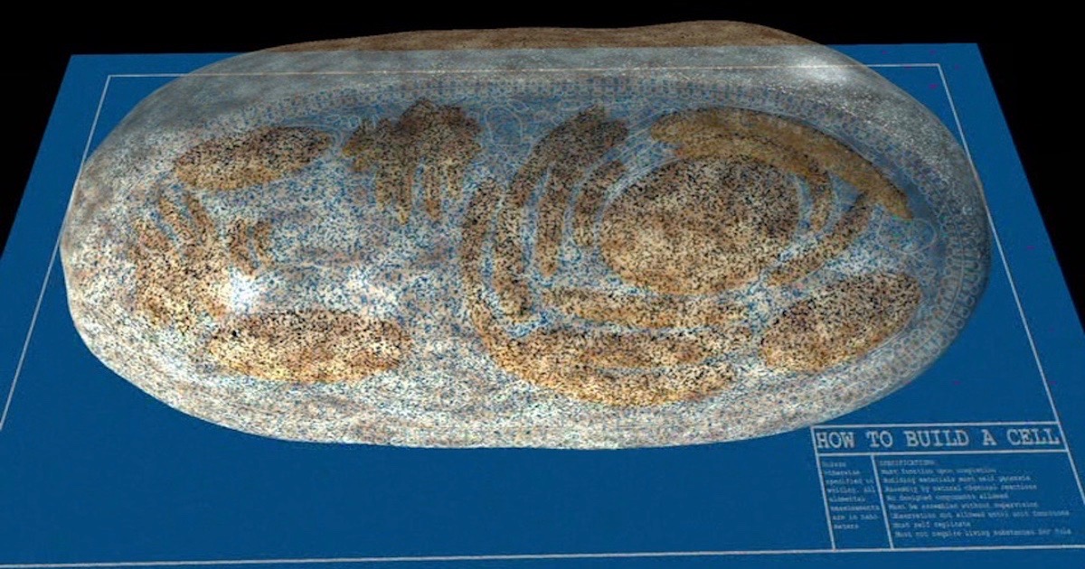
Image source: Illustra Media, Origin: Design, Chance, and the First Life on Earth.
